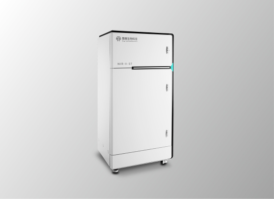
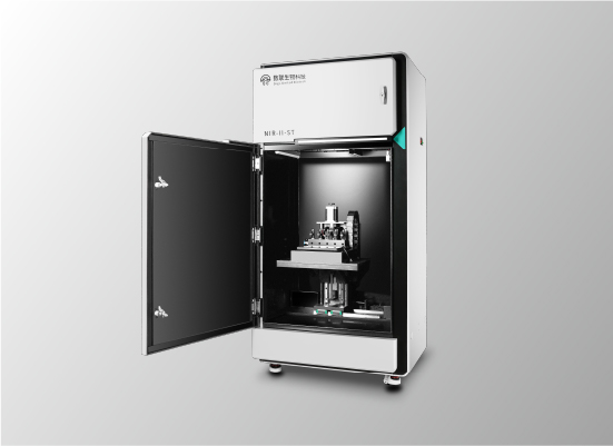
NIR-II-ST is a high-performance in vivo imaging system in the NIR-II window (900-1700 nm) for small animals, which can be paired with a variety of deeply cooled InGaAs cameras. The high-performance imaging lenses combined with 3D translation tables can achieve large and small field of view with high signal-to-noise ratios (2 cm×2 cm-20 cm×20 cm). The system is equipped with automatic operating platform and can be autofocused. In addition, the multi-channel small animal anesthesia system can realize imaging of multiple mice simultaneously. X-ray, near infrared LED and lasers with multi-wavelengths can be chosen as the light sources. Self-developed control and image processing software can make the operation simpler and image processing more comprehensive
Applications:
In vivo imaging of multiple organs; fluorescence guided surgery; in vivo vascular imaging, lymphatic imaging, tumor imaging; inflammation detection and monitoring, drug tracking, in situ disease detection, etc.
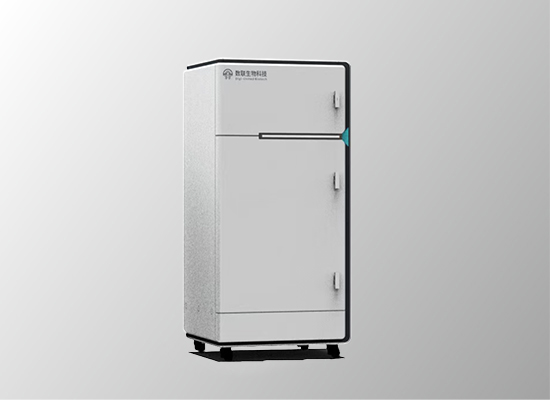
V-NIR-II covers the full spectrum region from visible/NIR-I/NIR-II (400-1700 nm). Due to the deeply cooled visible and InGaAs camera, the system has unbeaten signal-to-noise ratio with adjusted imaging field of view (2 cm×2 cm-20 cm×20 cm). The two cameras can be easily switched through the software. A three-dimensional translation stage (integrated with a automatic heating platform that can maintain the body temperature of anesthetized mice for a long time ) and other lasers, X-ray, and near-infrared LED are optional. In addition, the two camera lens of the system can be autofocused. The multi-channel small animal anesthesia system enables long time imaging. X-ray, near infrared LED and lasers with multi-wavelengths can be chosen as the light sources. Self-developed control and image processing software can make the operation simpler and image processing more comprehensive
Applications:
Whole-body and local imaging in live animals, fluorescence-guided surgery, vascular imaging, lymphatic imaging, tumor imaging, inflammation detection and monitoring, drug tracking, in situ disease detection, drug efficacy evaluation, bioluminescence, photothermal, photodynamic therapy, etc.

NIR-II-LT is a unique in vivo imaging system in the NIR-II window for characterizing long fluorescence lifetime of probes (microsecond to millisecond) in vivo or in vitro, with the the imaging field of view 2 cm×2 cm. The system is equipped with a deep cooling near infrared camera, which can realize long-time exposure for weak signal acquisition. The developed fluorescence lifetime software can acquire the sample signal, adjust the parameters and obtain the fluorescence lifetime data easily. In addition to the lifetime imaging mode, the system also has the function of wide-field fluorescence imaging, and the two imaging modes can be switched electrically through the software. A three-dimensional translation stage (integrated with a automatic heating platform) and other lasers, X-ray, and near-infrared LED are optional. A multi-channel small animal anesthesia system is also equipped and enables simultaneous imaging of multiple mice.
Applications:
Quantitative analysis of multi-component in vivo, immunohistochemical analysis in vivo, live cell labeling and quantitative analysis of tumor microenvironment in vivo
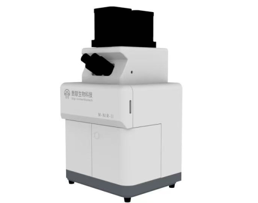
M-NIR-II is a new microscopic imaging system in the second near-infrared region with optimized and designed specifically optical paths. Its flexible modular enables the system to simultaneously use two or even three deeply cooled InGaAs cameras to achieve microscopic imaging in deep tissue. Besides, it also enables to dynamically trace a variety of different biomarkers in real time. Compared to traditional visible light microscopy, M-NIR-II has a much deeper penetration depth, higher resolution and signal-to-noise ratio. The systems can be equipped with multiple objective lenses with adjustable imaging field of view. It can also be connected with a variety of lasers with different wavelengths for multiple excitation.The multi-channel small animal anesthesia system can realize simultaneous imaging of multiple mice.
Applications:
Tumor imaging, microangiography, cell staining experiment, live cell tracing, immune cell interaction mechanism study, cancer cell migration, pathological section analysis, etc.
The V-ST series is a visible and NIR-I in vivo imaging system for small animals, covering the 300-1000nm spectral range. It enables bioluminescence and fluorescence imaging with high detection sensitivity. The system's multi-channel anesthesia module supports simultaneous imaging of multiple mice. With strong compatibility, its functionalities can be expanded to include full-spectrum live imaging, X-ray excitation modules, 3D imaging modules, thermal imaging modules, in-situ spectroscopy testing, etc.
NIR-vista is a miniaturized imaging system in the second near-infrared region with a compact design. The system takes up only a little space and facilitates installation and subsequent instrument movement. The system features high resolution (640×512), fast scanning speed (more than 250 frames per second), low dark noise and easy operation, making it a cost-effective imaging instrument. Multiple lasers of different wavelengths can be attached for multiple excitations, and the combined multi-channel small animal anesthesia system can realize simultaneous imaging of multiple mice.
Applications:
Suitable for studying of all kinds of in vivo processes that requiring fluorescence or bioluminescence signals. Examples include tracking tumor metastasis, angiography, pharmacokinetic studies, and evaluating the efficacy of new drugs.
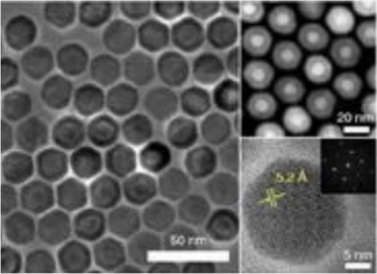
The morphology, size and multilayer core-shell structure of rare earth nanoprobes can be precisely synthesized. By changing the dopant ions, the absorption wavelengths (655 nm, 730 nm, 808nm, 860 nm, 980 nm, 1064 nm, 1208 nm, 1525 nm) and the emission wavelengths (1060 nm, 1155 nm, 1289 nm, 1475 nm, 1525 nm, 1632 nm, 1800 nm, etc.) can be regulated. In addition, the fluorescence lifetime of the nanoprobes can also be adjusted in the range of three orders of magnitude (microsecond - millisecond) for a single emission wavelength.
 xusy@digi-united.com
 +86 178 9511 3630


Design by DOHARD TEL: 0574 8808 1302
Copyright © 2023上海数联生物科技有限公司版权所有 | 沪ICP备2021012949号-1
